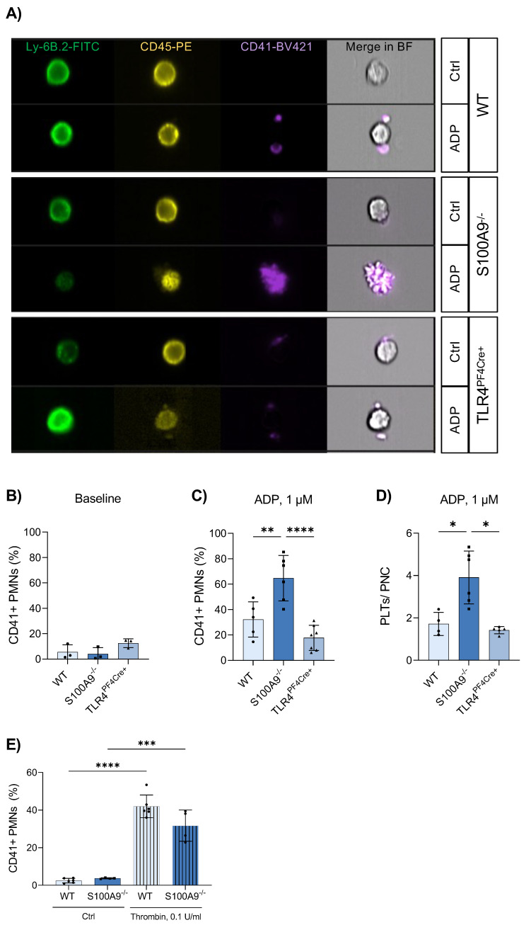
Figure 3.
S100A9 deficiency results in an increased number of platelet–neutrophil complexes. Whole-blood samples from healthy WT, S100A9−/− and TLR4PF4Cre+ mice were stimulated with 1 µM ADP or left untreated. Neutrophil–platelet complexes were analysed via imaging flow cytometry and identified as Ly-6B+CD45+CD41+ aggregates (A). The percentage of neutrophil–platelet complexes (CD41 + PMNs) was assessed under baseline conditions (B) and after ADP stimulation (C). The number of platelets per complex was determined manually (D). The formation of platelet–neutrophil complexes was simulated in vitro by coculturing platelets and bone marrow-derived neutrophils in media supplemented with 0.1 U/mL thrombin. CD41+ neutrophils (PMNs) were analysed via flow cytometry (E). Data are mean ± SD, n = 4–6, * p < 0.05, ** p < 0.005, *** p < 0.001, **** p < 0.0001, ordinary one-way ANOVA.

