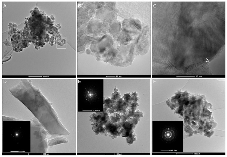
Figure 6.
Transmission Electron Microscopy (TEM) of gQ-n3 after dispersion in ultrapure water and probe sonication. Large agglomerates of 20–30 nm quartz nanoparticles are evidenced at low magnification (A). The particles in the highlighted white square are imaged at higher magnification (B). High-resolution image of a portion of the agglomerated quartz (C) highlights crystalline core, with several diffraction planes visible, and the amorphous external layer (indicated by the asterisk) formed during high-energy milling. A larger submicrometric highly crystalline quartz particle ((D), SAED in the inset). Nanometric agglomerates of milled quartz at low magnification and their corresponding large-field SAED evidenced multiple reflections and rings that indicate a nanometric size of the primary particles ((E,F), SAED in the inset).

