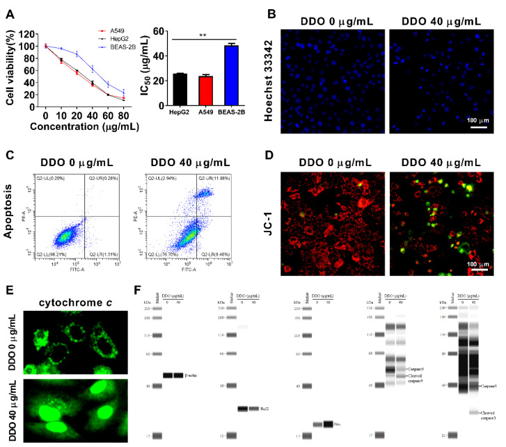
Figure 10.
Cytotoxicity and apoptosis-inducing effect of compound DDO. HepG2, A549, and BEAS-2B cells were treated with or without a series concentration of DDO for 24 h, then cell viability was determined by (A) CCK8 assay and IC50 was calculated; the values represent mean ± standard deviation of three independent experiments. A549 cells were treated with DDO (40 µg/mL) for 24 h, apoptosis cells were stained with (B) Hoechst 33342 and (C) annexin V-FITC/PI kit, and detected with ImageXpress Micro Confocal High-Content Imaging System and flow cytometry, respectively. (D) After being treated with DDO (40 µg/mL) for 24 h, A549 cells were stained with JC-1 to observe the mitochondrial membrane potential. A549 cells were treated with DDO (40 µg/mL) for 9 h, (E) a part of cells was used for immunofluorescence analysis of the release of cytochrome c from mitochondria, (F) and another part of cells was harvested to prepare total proteins, which were then subjected to Simple Wes System for detection of apoptosis-related proteins. The values represent mean ± standard deviation of three independent experiments, ** p < 0.01.

