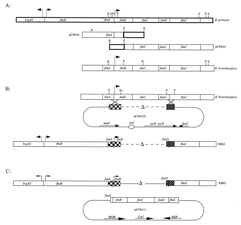
FIG. 1.
(A) Fragments of B. pertussis DNA homologous between B. pertussis and B. bronchiseptica were used to integrate a plasmid into the B. bronchiseptica genome. Flanking regions of B. bronchiseptica DNA were then isolated while the plasmid was excised from the genome. Thick-lined boxes represent the B. pertussis DNA and show the organization of the fim locus; thin-lined boxes represent B. bronchiseptica DNA. Two KpnI-PstI B. pertussis fragments were used to isolate B. bronchiseptica DNA cloned as plasmids pCMA4 and pCMA6. Restriction analysis of B. bronchiseptica DNA revealed differences between B. pertussis and B. bronchiseptica DNA as is represented by the absence of certain restriction sites in B. bronchiseptica. The asterisk represents extra DNA in B. bronchiseptica that does not correspond to B. pertussis sequences in this region. There is evidence that fimA in B. bronchiseptica may be a complete and functional gene (7). A, AlwNI; As, AspI; K, KpnI; N, NspI; P, PstI; S, SmaI; X, XmaI. (B) SalI-AspI and PstI-PstI fragments from B. bronchiseptica were ligated in frame to create the ΔfimBCD mutation. pCMA10 represents the allelic exchange plasmid used to introduce the deletion into the B. bronchiseptica chromosome. RB63 represents the genetic organization of the ΔfimBCD strain. (C) The minimal open reading frame of the fimbrial biogenesis operon (fimBCD) containing the intact fimB promoter was used to complement the Fim− mutant, RB63. pCMA11 represents the complementation plasmid.

