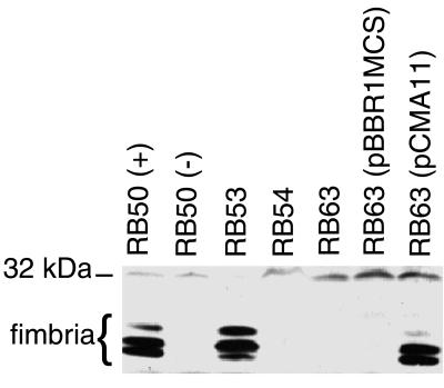
FIG. 2.
Western immunoblot analysis of whole-cell lysates of RB50 (+ phase), RB50 (− phase), RB53, RB54, RB63, RB63(pBBR1MCS), and complementation strain RB63(pCMA11). Approximately 25 OD600 units of lysates were loaded per lane and probed with a 1:4,000 dilution of anti-Fim3 antibody. The cluster of bands running at approximately 21 to 24 kDa represents fimbriae. The position of the 32-kDa molecular weight marker is shown on the left.

