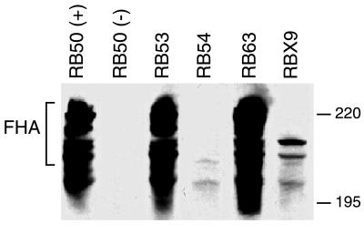
FIG. 3.
Western immunoblot analysis of whole-cell lysates of RB50 (+ phase), RB50 (− phase), RB53, RB54, RB63, and RBX9 probed with anti-FHA antibody. The cluster of bands running at approximately 220 kDa represents FHA. Positions of molecular weight markers (220 and 195 kDa) are shown on the right.

