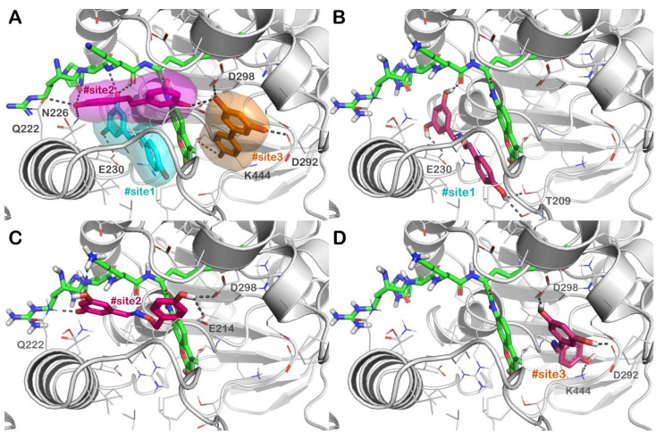
Figure 3.
(A) Crystal structure of SIRT1 (grey cartoon representation) in complex with resveratrol (sticks representation) (PDB ID 5BTR); the different binding sites of resveratrol, namely, #site1, #site2, and #site3 are represented in cyan, magenta, and orange, respectively; (B–D) binding mode of compound 3d (dark purple sticks) into SIRT1 with particular focus on #site1 (panel B), #site2 (panel C) and #site3 (panel D). The p53-AMC-peptide is reported as green sticks. Residues in the binding sites are represented by lines while hydrogen bonds are shown as grey dotted lines. Pictures were generated by PyMOL software (The PyMOL Molecular Graphics System, v1.8; Schrödinger, LLC, New York, NY, USA, 2015).

