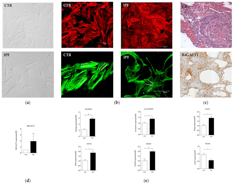
Figure 2.
In Vitro characterization of IPF primary cell line. (a) Primary culture of human normal fibroblast (upper) and IPF (lower) (×10 magnification). (b) Representative bright field images on actin of normal fibroblast and IPF primary cell cultures (×40 magnification). (c) H&E corresponding to lung biopsy of the patient with UIP from which the primary cell culture was isolated and IHC analysis (×20 magnification). (d) mRNA expression level of B4GALT1. (e) mRNA levels of stemness genes and EMT genes between normal fibroblast and IPF verified by qRT-PCR. The data are expressed as mean ± SD.* p < 0.05; ** p < 0.005.

