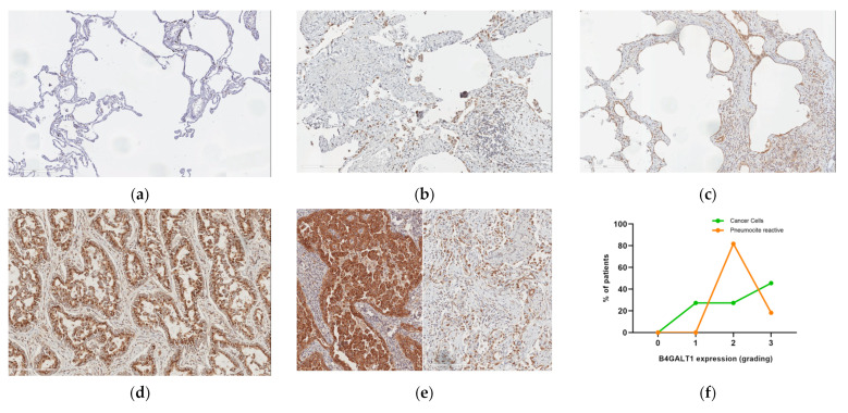
Figure 3.
B4GALT1 expression in lung fibrosis and lung cancer (×20 magnification). (a) An example of near-normal lung tissue is shown (B4GALT1 staining); (b) positive staining in lung cancer; (c) tumor (on the left) and UIP (on the right) of the same patient show simultaneous strong cytoplasm B4GALT1 expression; (d) strong B4GALT1 expression is observed in most fibroblastic foci in UIP both in connective tissue and alveolar epithelium; (e) and possibly in UIP fibrous reactive remodeling of lung tissue not related to IPF; (f) quantification of grading expression of B4GALT1; (g) there are B4GALT1-positive immune lymphocyte infiltrating cells in neoplastic and UIP tissue samples; and (h,i) capillary endothelium expressing B4GALT1 was observed, in the same patient, both in the immediately peritumoral connective tissue area and adjacent to the UIP pattern fibrous.


