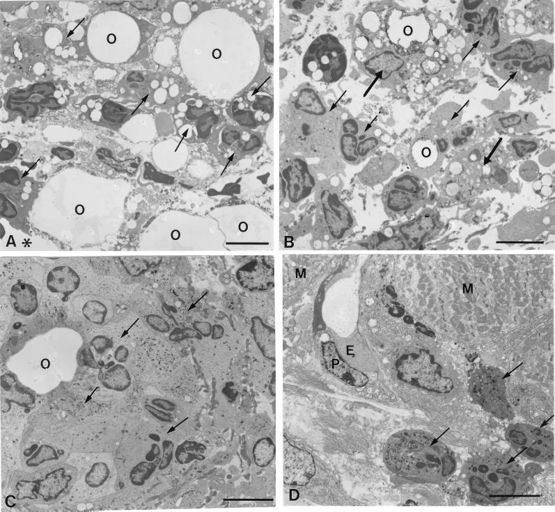
FIG. 5.
Electron micrographs of infiltrating cells in the pouch wall treated with Rhodococcus sp. strain 4306 TDM at 1 (A) or 3 (B to D) days after injection. At day 1, the necrotic tissue facing the pouch cavity (asterisks) contained many neutrophils (arrows). (A) Neutrophils were attached to the oil droplets and phagocytosed small droplets. (B) At day 3, a few macrophages (thick arrows), which phagocytosed the oil droplets, joined the neutrophil infiltrate (arrows) in the necrotic tissue. (C) In the granulomatous tissue, neutrophils accumulated around the oil droplets. (D) Beneath the muscular layer, new vessels extended, being accompanied by neutrophil infiltrate (arrows). E, endothelial cells; P, pericytes; O, oil droplets; M, muscular layer. Bar, 5 μm.

