Figure 1.
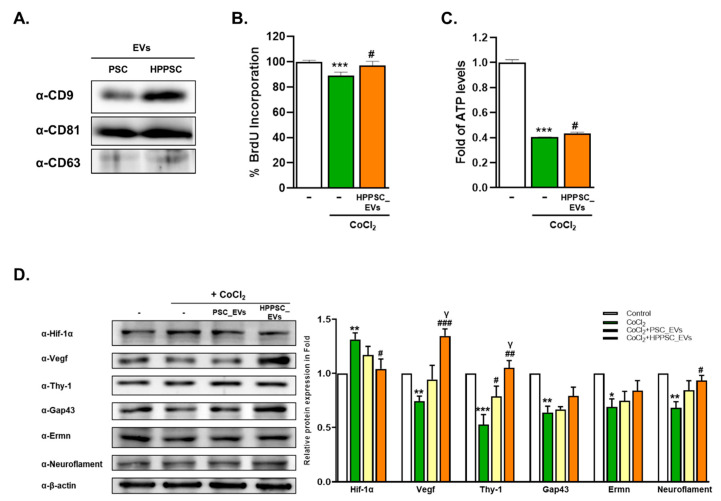
Characterization and recovery effects of hHPPSCs-derived EVs. (A) Isolated EVs expressing classic exosomal markers CD9, CD81, and CD63. R28 cells were treated with CoCl2 (200 μM). After incubation for 9 h, the cells were treated with EVs. (B) BrdU assays performed after 24 h. Data are presented as a mean ± SEM. (C) Determination of ATP production was presented as a fold (mean ± SEM). (D) Western blot analyses of target protein expression levels, using R28 lysates with CoCl2. The levels of protein expression are quantified (bottom-panel). Significantly difference was estimated using an unpaired t test (* p < 0.05, ** p < 0.005, *** p < 0.001 vs. the control; # p< 0.05, ## p< 0.01, ### p < 0.005 vs. CoCl2; γp < 0.05 vs. PSC_EVs). All experiments were performed in triplicate.

