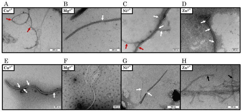
Figure 11.
Negatively stained TEM micrographs of (A–D) C-TERb and (E–H) C-TERa (CC-TER = 5 µM) incubated for 144 h (6 days) in TBS at ~20 °C in the presence of (A,E) Cu2+, (B,F) Mg2+, (C,G) Ni2+, and (D,H) Zn2+ cations (CM2+ = 100 µM). Under certain conditions, the formed fibrils present breaking points (indicated by red arrows) and happen to be fragmented (breakages are indicated by white arrows). In Panel H, black arrows pinpoint single straight fibrils (SSFs) that cluster into multiple straight fibrils (MSFs). The scale bar is provided at the bottom right of each TEM micrograph.

