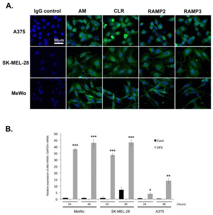
Figure 2.
Depiction of the extent to which AM signaling is expressed and regulated in melanoma cells. (A) AM and receptors expressed in melanoma cells depicted using immunofluorescence of A375, SK-MEL-28, and MeWo cells stained with antibodies against AM, CLR, RAMP2, and RAMP3, revealing localization in the cytoplasm. Negative control for immunostaining was achieved with IgG-control. (B) AM expression induced by a hypoxia mimetic in melanoma cells. Total RNA (1 μg, DNA free) prepared from MeWo, SK-MEL-28, and A375 cells were reverse transcribed into cDNA under normoxic or hypoxic conditions and relative AM mRNA was estimated using a real-time quantitative reverse transcriptase polymerase chain reaction. There were significant differences between cells treated with desferrioxamine mesylate (DFX) and untreated control cells in terms of AM expression: * p < 0.05; ** p < 0.01; *** p < 0.001. Each experiment is representative of five independent experiments. Results are shown as means ± SD.

