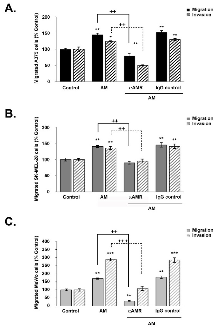
Figure 4.
AM regulates melanoma cell migration and invasion in vitro. (A–C) The bottom wells of all chambers were filled with DMEM for A375 and SK-MEL-28 cells or MEM for MeWo cells containing 2% fetal bovine serum in the presence of control buffer (control) or AM (10−7 M). A375 ((A), 2 × 10⁴ cells), SK-MEL-28 ((B), 2 × 10⁴ cells), or MeWo ((C), 1 × 10⁵ cells) cells pretreated for 30 min with αAMR (70 μg/mL) or control IgG (70 μg/mL) were placed in the upper chamber and incubated for 16 h at 37 °C. The cells that migrated were stained with 4′, 6′-diamidino-2-phenylindole and counted at 50x magnification using a microscope. Data are expressed as the number of migrated cells in 10 high-power fields, and the values represent the mean ± SD of four independent experiments, each performed in triplicate. The asterisk (*) is used for comparison to control cells (* p < 0.05; ** p < 0.01; *** p < 0.001) and the plus symbol (+) is used for comparison to AM-treated cells (++ p < 0.01; +++ p < 0.001).

