Figure 5.
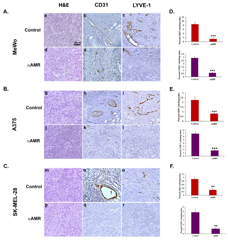
Analysis of in vivo Matrigel plug bioassays indicates that AM secreted by melanoma cells induces angiogenesis and lymphangiogenesis. (A–C) A total of 0.8 mL of growth factor-depleted Matrigel was admixed to MeWo ((A), 1 × 10⁶ cells) (a,b,c,d,e,f), A375 ((B), 1.5 × 10⁶ cells), or SK-MEL-28 ((C), 2 × 10⁶ cells) cells and administered to C57BL/6 mice via s.c. injection at the abdominal midline. Administration of αAMRs or control IgG was conducted intraperitoneally every three days (starting 24 h after initial Matrigel injection and for 15 days thereafter) in C57BL/6 mice. Formalin was used to fix Matrigel plugs, which were then embedded, sectioned, and used for immunohistochemical analysis. Figure 5A–C depict microphotographs of histochemical-stained Matrigel sections for H & E (a,d,g,j,m,p), blood vessel staining with the CD-31 antibody (b,e,h,k,n,q), and lymphatic vessels with the anti-LYVE-1 antibody (c,f,i,l,o,r) derived from Matrigel plugs mixed with melanoma cells treated with either αAMRs or control IgG. Each panel represents multiple fields, including five plugs in each group. Scale bar, 50 μm. (D–F) Quantitative assessment of cell density for CD31- and LYVE-1-positive cells as assessed by staining conducted on the entire surface of the corresponding slides using CALOPIX software. (v2.10.16 by Tribvn) MBF_Image J 1.52a software was used for the analysis. The values represent the means ± SD (** p < 0.01; *** p < 0.001).

