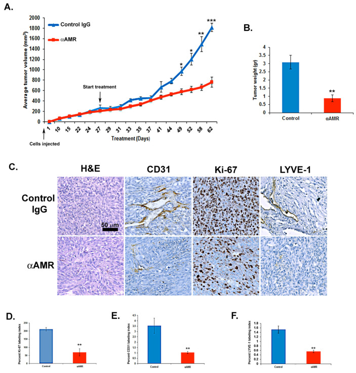
Figure 7.
AM signaling blockade inhibited the growth of MeWo xenografts in vivo. (A) MeWo cells (2 × 10⁶) were injected subcutaneously into the flanks of athymic nude mice (6 weeks old) (n = 10 in each group). Mice with tumor volumes averaging 250 ± 50 mm3 received intraperitoneal injections of αAMRs (12 mg/kg) every 3 days. Control mice were treated with 12 mg/kg of nonspecific isotype control immunoglobulin G (IgG). Measurements of tumor volume demonstrate differences in the growth of animals treated with αAMRs (n = 10) and control IgG (n = 10) during the 52-day schedule, * p < 0.05; ** p < 0.01; *** p < 0.001. (B) Tumors were weighed immediately after excision and the average tumor is indicated as the mean ± SD (n = 10), ** p < 0.01. (C) αAMRs-treated tumors are less vascular and depleted of vascular and lymphatic endothelial cells. LYVE-1, CD31, and Ki-67 antibodies and hematoxylin and eosin were used to stain the tumor sections. The figure depicts Ki-67 positive cells, with each section analyzed using 10 magnification fields (400×). Microvessel density was determined using immunohistochemical staining of the CD-31 marker of the endothelial cell surface. The density of cells staining positive for Ki-67 (D), CD-31 (E), or LYVE-1 (F) was assessed quantitatively based on the entire slide surface using CALOPIX Software v2.10.16 by Tribvn. Analysis was conducted with MVF_Image J1.52a software. The values shown represent the means ± SD, ** p < 0.01.

