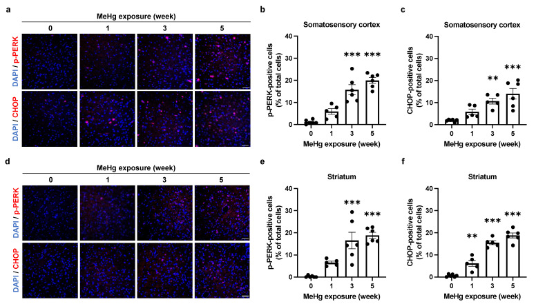
Figure 4.
Effect of MeHg exposure on PERK branch in the mouse brain. (a,d) Detection of p-PERK and CHOP in the somatosensory cortex (a) or striatum (d) of ERAI-Venus mice exposed to MeHg for the indicated times. Each scale bar represents 50 μm. (b,e) Quantification of p-PERK-positive cells in the somatosensory cortex (b) or striatum (e). (c,f) Quantification of CHOP-positive cells in the somatosensory cortex (c) or striatum (f). Data are expressed as the mean ± S.E.M. values (n = 5–6, ** p < 0.01, and *** p < 0.001: significant difference compared with control mice without MeHg exposure (0 weeks) by one-way ANOVA with Dunnett’s post hoc test).

