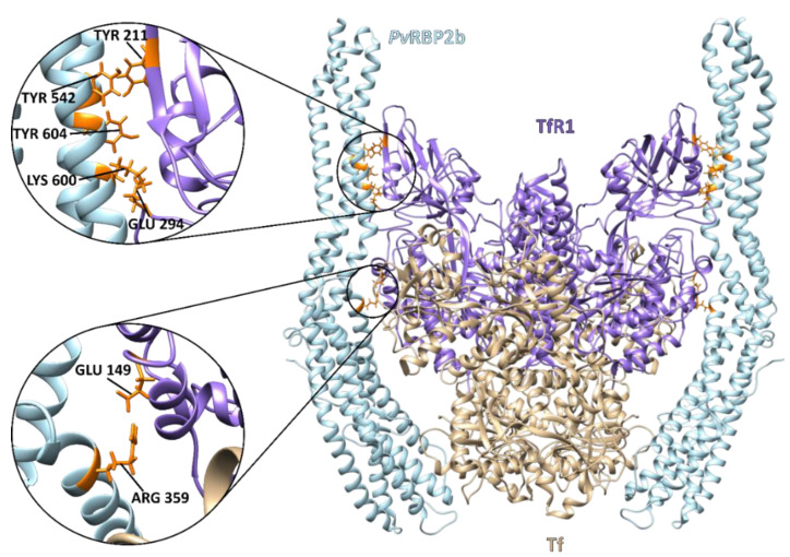
Figure 3.
Cryo-electron microscopy structure of the ternary PvRBP2b complex bound to human TfR1 and Tf (PDB: 6D04). The complex consists of homodimer TfR1 (residues 120–760) (purple) bound to two molecules of iron-loaded Tf (residues 1–679) (beige) and two molecules of PvRBP2b (residues 168–633) bound on either side (light blue). The critical PvRBP2b and TfR1 binding residues are enlarged and highlighted in orange [40].

