Figure 1.
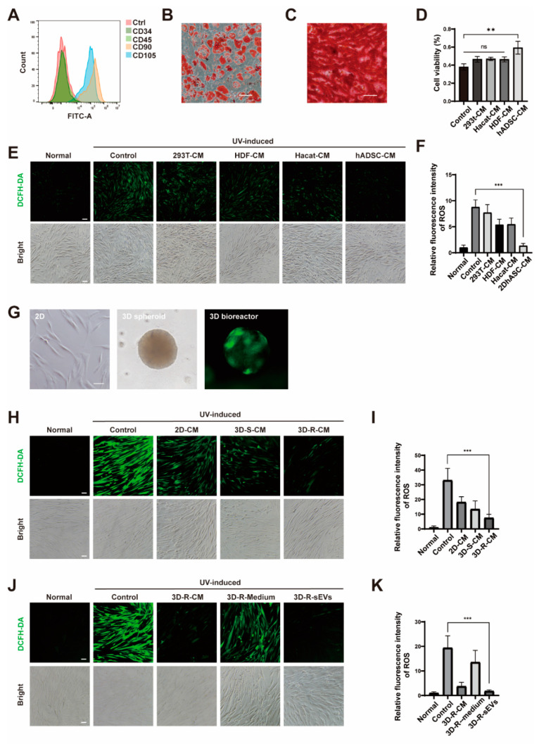
3D-R-sEVs has a significant anti-photoaging effect. (A) Flow cytometry analysis of CD34, CD45, CD90, and CD105 expression on hADSCs surface. (B) Oil Red O stain of adipo-differentiated hADSCs. Scale bar: 200 μΜ. (C) Alizarin Red stain of osteo-differentiated hADSCs. Scale bar: 200 μΜ. (D) showed the viability of HDF cells which pre-incubated with the conditioned medium (CM) of HEK-293T cells, HDF, HaCaT, and hADSCs, respectively, for 24 h after being exposed to 10 J/cm2 dose of UVA tested by CCK-8 (n = 3). (E,H,J) Representative images showed the level of ROS (green) in different treatment groups of HDF cells. Scale bar: 100 μΜ. (F,I,K) Relative quantitative analysis of fluorescence intensity of ROS in different treatment groups of HDF cells (n = 10). One-way ANOVA and Tukey post hoc test analyses were performed. Error bar, mean ± SD. ns represents not significant, ** p ≤ 0.01, *** p ≤ 0.001. (G) Morphology of hADSCs in 2D, 3D spheroid, and 3D bioreactor culture, respectively. Scale bar: 100 μΜ.

