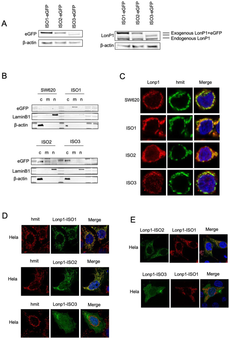
Figure 2.
Cellular localization and distribution of three isoforms of Lonp1. (A). Immunoblot detecting Lonp1 using anti eGFP (left panel) and anti Lonp1 (right panel) Abs in cells transfected with Lonp1 ISO-eGFP, ISO2-eGFP, or ISO3-eGFP. The position of the bands corresponding to the exogenous, eGFP-tagged form and the endogenous form of Lonp1 is indicated. (B). Representative immunoblots showing Lonp1 expression in cytosolic (C), mitochondrial (M), and nuclear (N) fractions in SW620 cells transfected with constructs bearing Lonp1 isoforms tagged at C-term with enhanced green fluorescent protein (eGFP). Lonp1 has been detected using anti-eGFP antibody. (C). Representative confocal microscopy images of SW620 cells transducted with three different constructs bearing Lonp1 isoforms tagged at C-term with enhanced green fluorescent protein (eGFP), namely Lonp1-ISO1, Lonp1-ISO2, or Lonp1-ISO3. Mitochondria were stained with anti-hMit and nuclei were counterstained with DAPI. (D). Representative confocal microscopy images of HeLa cells transfected with Lonp1-ISO1, Lonp1-ISO2, or Lonp1-ISO3. Mitochondria were stained with anti-hMit and nuclei were counterstained with DAPI. (E). Representative confocal microscopy images of HeLa co-transfected with constructs bearing Lonp1 isoforms tagged with enhanced green fluorescent protein (eGFP, in green) or mCherry (in red). Nuclei were counterstained with DAPI.

