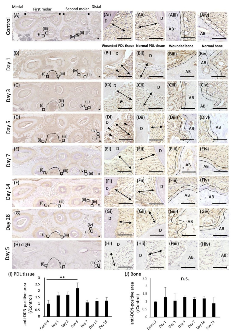
Figure 1.
Expression of DCN in wounded rat periodontal tissue. Immunochemical analysis of DCN expression was performed in wounded rat periodontal tissue, including PDL tissue and the alveolar bone. Horizontal sections of rat maxilla were prepared, and preoperative sections were used as a control (A). Localization of anti-DCN antibody-positive areas in the wound site was examined on days 1 (B), 3 (C), 5 (D), 7 (E), 14 (F), and 28 (G). Magnified images of wounded PDL tissue (i), normal PDL tissue (ii), wounded alveolar bone (iii), and normal alveolar bone (iv) in each section are shown. Control IgG was used for the negative control (H). Nuclei were counterstained with hematoxylin. Arrowheads indicate anti-DCN antibody-positive areas. Double arrows indicate PDL tissue. The inside of the two dotted lines exhibits osteoblastic layers. D, dentin; AB, alveolar bone. Bars = 100 µm. (I,J) Graphs show the quantification of anti-DCN antibody-positive areas in the PDL tissue (I) or the osteoblastic layer around the alveolar bone (J). Anti-DCN antibody-positive areas in preoperative periodontal tissue were used as the control. Values are shown as the fold increase relative to the control. ** p < 0.01, n.s. = no significance, n = 3.

