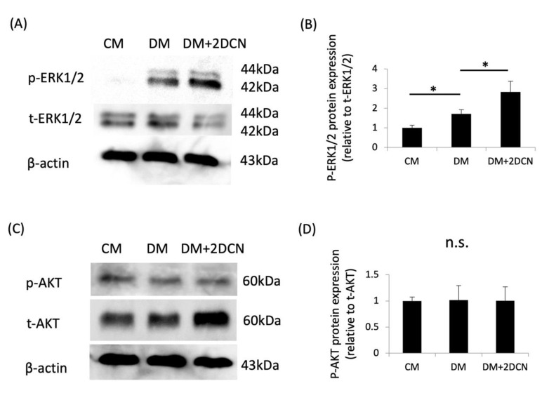
Figure 4.
Expression of phosphorylated-ERK1/2 and phosphorylated-AKT during the DCN-induced osteoblastic differentiation of HPDLSCs. HPDLSCs were cultured in 10% FBS/α-MEM (CM), CM with 1.5 mM CaCl2 (DM), or DM on a 2 μg/mL DCN coating (DM + 2 DCN) for 15 min. (A) Western blot analysis was performed to determine the expression of phosphorylated-ERK1/2 (p-ERK 1/2) and total-ERK1/2 (t-ERK 1/2). β-actin was used as a loading control. (B) The graph shows the quantification of p-ERK 1/2 expression. Normalization of protein expression was performed against t-ERK1/2 expression. (C) Western blot analysis was performed to determine the expression of phosphorylated-AKT (p-AKT) and total-AKT(t-AKT). β-actin was used as a loading control. (D) The graphs show the quantification of p-AKT expression. Normalization of protein expression was performed against t-AKT expression. Values are the mean ± SD of three indepenDent. experiments. * p < 0.05, n.s. = no significance.

