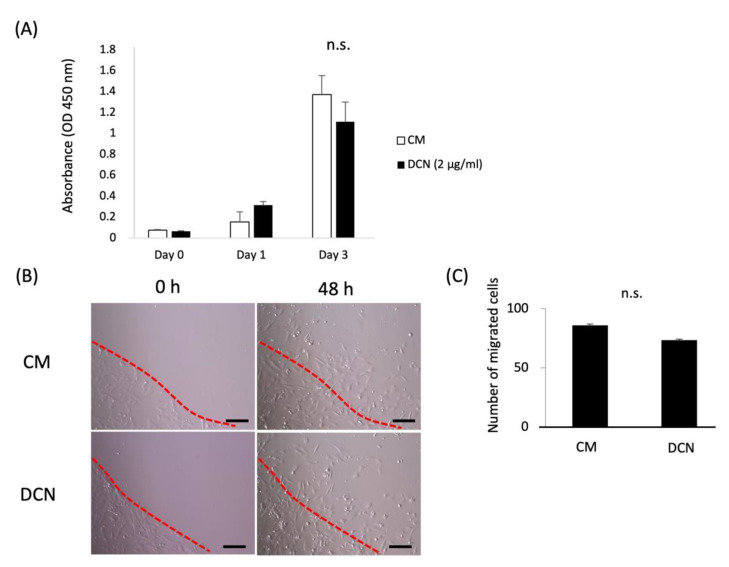
Figure 5.
Effects of DCN on the proliferation and migration of HPDLSCs. (A) HPDLSCs were cultured in 10% FBS/α-MEM (CM) or CM on a 2 μg/mL DCN coating (DCN) for 0, 1, and 3 days. A proliferation assay was performed using the WST-1 proliferation assay kit at an absorbance of 450 nm. (B) Migration of HPDLSCs was analyzed by a ring cell migration assay. HPDLSCs were cultured in CM or DCN for 48 h, and then migrated cells were counted. Dotted lines delineate the ring edges. Bars = 200 µm (C) The graph shows the quantification of migrated cells. Values are the average of three random fields per well. n.s. = no significance.

