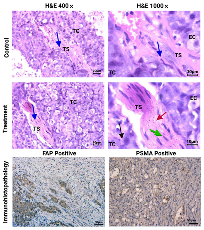
Figure 6.
Histopathological analysis (H&E staining). Light micrographs of representative sections at 400× and 1000× magnification of HCT116 tumors from [177Lu]Lu−iFAP/iPSMA-treated and untreated (control) mice. Cancer-associated fibroblasts (CAFs) can be observed in the tumor stroma (TS) (blue arrow). Note nanoparticle clusters of [177Lu]Lu−iFAP/iPSMA in TS (red arrow), in CAFs (green arrow), and in tumor cells (TC) (black arrow), but not found in endothelial cells (EC). PSMA and FAP expression in HCT116 tumors (dark brown color) (200× magnification of) is also shown (immunohistopathology results).

