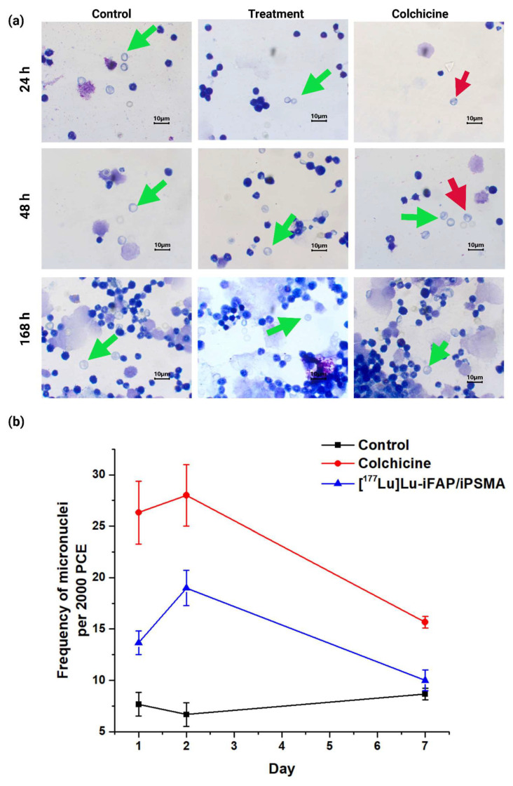
Figure 7.
(a) Light micrographs (Giemsa-Wright staining at 1000× magnification of) of bone marrow cells from healthy mice at 24, 48, and 168 h after i.v. administration of [177Lu]Lu−iFAP/iPSMA nanoparticles (treatment), colchicine (1 mg/kg) (positive control), and untreated mice (negative control). Green arrows show polychromatic immature erythrocytes, and red arrows indicate polychromatic erythrocytes (PCE) with micronuclei. (b) Micronucleus count values in immature bone marrow erythrocytes at 1, 2, and 7 d. Data are presented as mean ± standard deviation of three independent experiments. Non-significant difference (p > 0.05) between control and treatment at 7 d.

