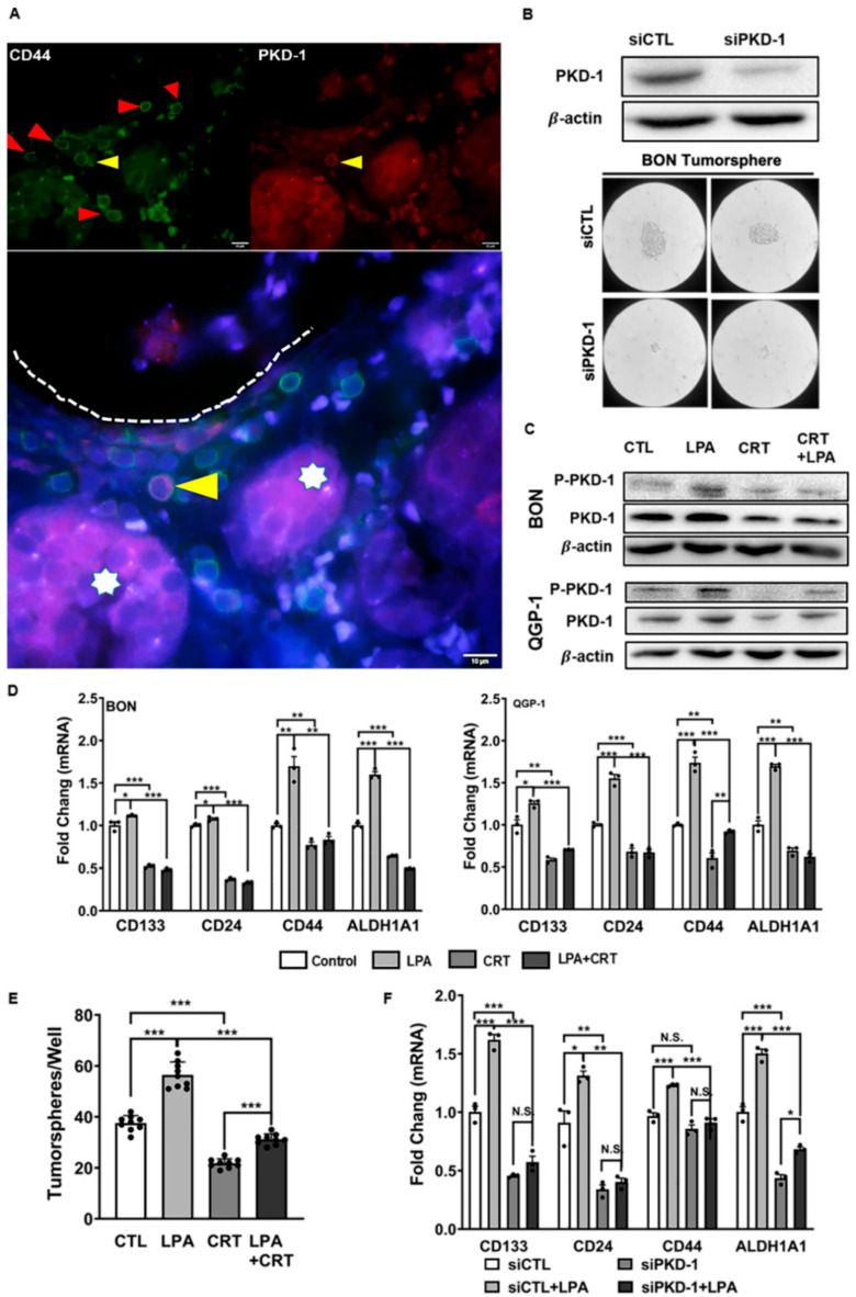
Figure 2.
PKD1 signaling in the maintenance of cancer stem-like features in pNETs. (A) Distribution of PKD1+ and CD44+ CSCs within the vascular niche. Human pNET specimens were co-stained with CD44 and PKD1 antibodies, followed by appropriate secondary antibodies, with DAPI staining the nuclei (Blue). Stem-like cells with CD44-positive (green), PKD1-positive (red) or both positive (pink) were observed under a fluorescence microscope. A few CD44-positive cancer stem-like cells tended to accumulate near the vascular lumen (red arrow heads). Cancer cell with moderate expression of both PKD1 and CD44 might be leaving tumor nests (stars) for the vascular lumen. The fluorescence images were acquired by a fluorescence microscope equipped with a CCD camera. Shown are representative images. Scale bar = 10 µm. (B) BON cells were transfected with siRNA control and siPKD1 to knock down PKD1. Knockdown efficiency was confirmed by Western Blots (upper panel). The control and BON cells with PKD1 knockdown were subjected to tumorsphere formation assays. Images were acquired by the OLYMPUS CK30 microscope. Representative images are shown for tumorsphere formation (lower panel). Scale bar = 200 µm. (C) Cell lysates were extracted from BON and QGP-1 cells exposed to the vehicle control, 10 μM LPA, 2 μM CRT0066101, or their combinations after 24 h. The expression levels of phosphorylated PKD1 and total PKD1 were detected by Western blots. Shown are representative images of triplicate experiments in BON (upper panel) and QGP-1 (lower panel) cells. (D) BON and QGP-1 cells were exposed to 10 μM LPA, 2 μM CRT0066101, or their combination for 24 h, and total RNA was extracted for the detection of mRNA levels of genes related to stemness properties by RT-qPCR. (E) Effect of PKD inhibitor in tumorsphere formation. BON cells were cultured in complete MammoCult™ medium with the treatment of 10 μM LPA, 2 μM CRT0066101, or their combination for 7 days. The number of mammary spheres was counted under the OLYMPUS CK30 microscope. (F) Control and BON cells with PKD1 knockdown were exposed to 10 μM LPA, 2 μM CRT0066101, or their combination for 24 h, and total RNA was extracted for the detection of mRNA levels of genes related to stemness properties by RT-qPCR. Triplicate experiments were performed, and the results are shown as the mean ± SEM. * p < 0.05, ** p < 0.01, and *** p < 0.001.

