Figure 2.
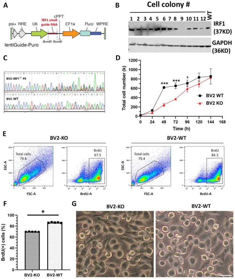
IRF1 knockout (KO) defers BV2 microglial proliferation. (A–C) IRF1 KO from BV2 microglial cells with CRISPR-Cas9 gene-editing technique. (A) The diagram depicting the insertion of IRF1 small guide (sg) RNA in the lentiviral plasmid. (B) The WB results showing that single-cell colony #9 is absent of IRF1 protein compared with the other cell colonies and wild-type (WT) cells. (C) DNA sequence showing the mutation of cell colony #9′s gene loci compared to the BV2-WT gene loci. (D–F) IRF1-KO defers the microglial cell proliferation rate. (D) Cell counts during BV2 cell growth. (E) Flow cytometry shows total live BV2 cells (left in the enclosed areas) and BrdU (+) BV2 cells (right in the boxed areas) for IRF1-KO BV2 (BV2-KO) and BV2-WT counterparts. (F) BrdU (+) cell numbers (%). n = 6. * p < 0.05; *** p < 0.001. (G) Cell morphological differences of BV2-KO and BV2-WT cells. Scale bar: 20 µm.

