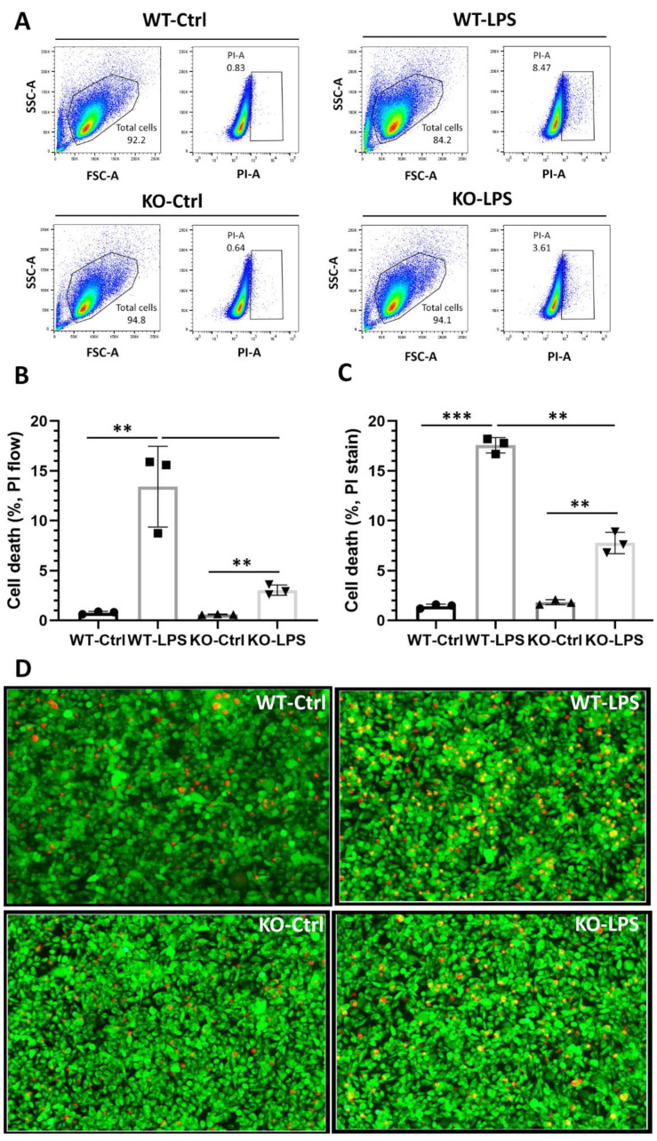
Figure 6.
IRF1 deletion reduces microglial cytotoxicity to photoreceptors. (A) PI-gated flow cytometry analysis for the cone-like 661 W cells treated with PBS control (WT-Ctrl), the conditioned media from wild-type microglia with LPS (WT-LPS), the conditioned media from IRF1-KO microglia with PBS control (KO-Ctrl), and the conditioned media from IRF1-KO microglia with LPS (WT-LPS). Total live cells were shown in the enclosed areas of left panels. PI-positive dead cells were shown in the boxed areas in the right panels (B) Cell death quantification by flow cytometry analysis. (C) Cell death quantification of the calcium AM and PI stained cone-like 661 W cells images. (D) The representative images showing the Calcium AM (green) and PI (red) stained cone-like 661 W cells. 40× magnification. The replicate numbers = 3. ** p < 0.01; *** p < 0.001.

