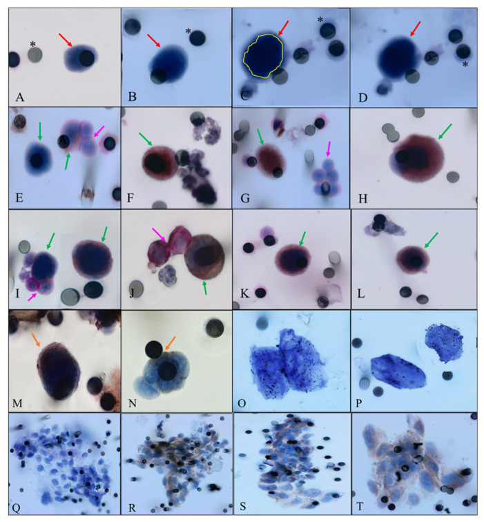
Figure 1.
Representative images. Green arrows indicate hybrid cells, pink arrows represent leukocytes, red arrows represent CTCs, orange arrows indicate hybrids cells stained with CD45, yellow circle indicate cell nucleus and asterisks represent membrane pore. (A,B) CTCs without any staining, visualized with hematoxilin. (C,D)The same CTC without any staining, in (C), nuclei defined by yellow line. (E) Hybrid cells. One positive for MC1-R (brown membrane) and one in a microemboli, stained with CD45 with two leukocytes (stained with CD45, visualized by permanent red). (F) Hybrid cell positive for MC1-R (brown membrane). (G) Hybrid cell positive for EpCAM (brown membrane) and a cluster of leukocytes (stained with CD45, visualized by permanent red). (H) Hybrid cell double positive for EpCAM (brown membrane) and MC1-R (visualized by permanent red). (I) Two hybrid cells. One visualized alone double stained with EpCAM and CD45 (brown and red) and the other surrounded by three leukocytes stained with CD45 (visualized by permanent red). (J) Hybrid cell stained with MC1-R, visualized by DAB and two leukocytes (stained with CD45, visualized by permanent red). (K,L) Hybrid cells positive for MC1-R/CD45 (brown and red membrane). (M,N) Hybrids cells stained with CD45 in just one side of the cytoplasmatic membrane, visualized by DAB. (O,P) CTCs positive for CEN8, indicating the presence of polyploidy. (Q) Microemboli visualized with hematoxylin. (R–T) Microemboli from patient positive for MC1-R (brown membrane). Images were taken at ×400 and ×600 magnification using a light microscope (Research System Microscope BX61—Olympus, Tokyo, Ja-pan) coupled to a digital camera (SC100—Olympus, Tokyo, Japan).

