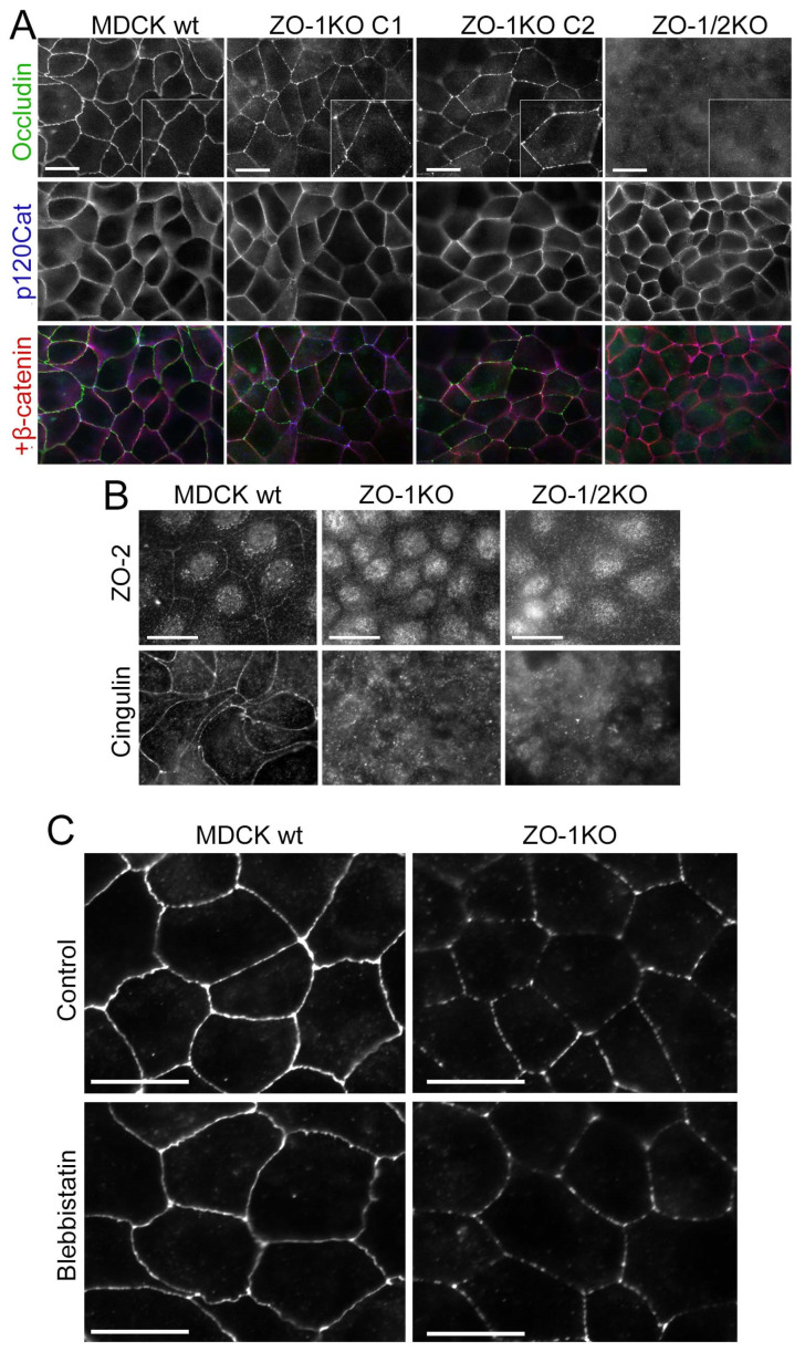
Figure 7.
ZO-1 and ZO-2 have distinct functions in TJ assembly. Analysis of wild-type and knockout MDCK cells seeded on glass and grown to confluence for 5 days by epifluorescence microscopy. Insets show higher magnifications to illustrate the discontinuous staining of occludin (A,B) Cells were stained for the junctional proteins. Note, occludin remained discontinuously distributed and recruitment of cytosolic TJ proteins was strongly attenuated. (C) Wild-type and ZO-1KO MDCK cells were treated with blebbistatin and then fixed and stained for occludin. Note, the distribution of occludin remained discontinuous in blebbistatin-treated ZO-1KO cells, indicating that the disrupted occludin arrangement was not due to the increased myosin activity. Magnification bars, 20 µm.

