Figure 4.
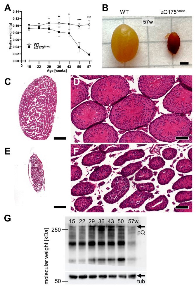
Male zQ175Δneo mice had significantly smaller testicles compared to WT littermates. Testicles of WT and zQ175Δneo mice were collected at 7-week intervals and weighed freshly right after perfusion. (A) Testicles’ weight was measured between 15 and 57 weeks of age of male WT (○) and zQ175Δneo (●) mice. Data are presented as mean ± SD; N = 6. Significance was calculated using Mann–Whitney test with Bonferroni-Dunn’s multiple comparisons test and is given as *: p ≤ 0.1, **: p ≤ 0.01 and ***: p ≤ 0.001. (B) Representative image showing a direct comparison of testicles of 57-week-old WT (left) and zQ175Δneo (right). The scale bar indicates 2 mm. (C–F) Representative images of H&E-stained sagittal testicular sections; overview (C,E; the scale bar indicates 500 µm) and close-up (D,F; the scale bar indicates 50 µm) of testicular seminiferous tubes in WT (C,D) and zQ175Δneo mice (E,F) at 57 weeks of age. (G) Western blot analysis of muHTT [polyQ (pQ) antibody, >>250 kDa] in testicles of zQ175Δneo mice 15–57 weeks of age. muHTT accumulates with increasing age (15–43 weeks) and then declines (50–57 weeks). Highly atrophic, 57-week-old (57w) testis have strongly reduced muHTT. The polyQ antibody (clone MW1) also detects N-terminal Htt fragments. Loading control: tub—β-tubulin, 55 kDa.

