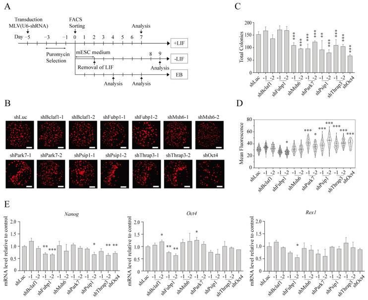
Figure 3.
The effect of knockdown of identified proteins on self-renewal and pluripotency of mESCs. (A) Experimental scheme of pluripotency and differentiation analyses. EB5/ReKO cells were transduced with retroviral vector expressing each shRNA followed by puromycin selection for 2 days. Five days after transduction, 1000 cells/well were sorted to a flat-bottom 96-well plate for culture in mESC medium with Leukemia Inhibitory Factor (+LIF) or without (-LIF) LIF, or 1 × 10³ cells/well to V-bottom 96-well plate for embryoid body (EB) formation (EB). (B) Whole well fluorescence images of hKO(+) mESC colonies cultured with LIF. EB5/ReKO cells treated with indicated shRNA and cultured in mESC medium with LIF. Whole well images were collected 7 days after sorting. Representative images are shown. Scale bars, 1500 µm. (C) Colony numbers of mESC treated with shRNA. Colony numbers were counted from the images collected in (B). Data represent the mean ± SEM from five independent experiments. ** p < 0.01, *** p < 0.001 versus EB5/ReKO cells treated with control shRNA (shLuc). (D) Mean fluorescent intensity of mESCs treated with shRNA. hKO fluorescent intensity in each colony was measured from the images collected in (B). Data represent the mean ± SEM from total colonies in each shRNA. * p < 0.05, *** p < 0.001 versus EB5/ReKO cells treated with control shRNA (shLuc). (E) mRNA level of pluripotency-related genes. Nanog, Oct4, or Rex1 mRNA levels in the EB5/ReKO cells prepared as (B) were determined 7 days after cell sorting. Data represent the mean ± SEM of three independent experiments. * p < 0.05, ** p < 0.01, *** p < 0.001 versus EB5/ReKO cells treated with control shRNA (shLuc).

