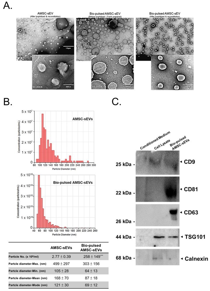
Figure 2.
Characteristics and advantages of bio-pulsed AMSC-sEVs. (A) Images of sEVs were obtained through transmission electron microscopy. Morphology did not alter after lyophilizing and reconstituting. (B) The particle size distribution and number were analyzed by the tunable resistive pulse sensing measurement system. (n = 3, data are expressed as the mean ± standard deviation; *** p < 0.001, compared with the AMSC-sEVs). (C) The extracellular vesicle markers of CD9, CD63, CD81, TSG101, and a contamination marker, calnexin, were examined through Western blot.

