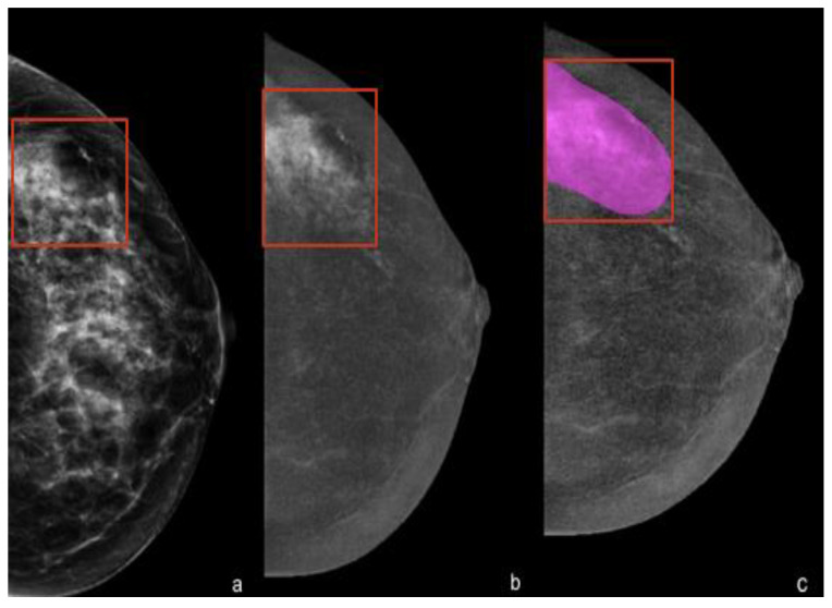
Figure 3.
Delineation of contours showing marked enhancement. (a) Left cranio-caudal full-field digital mammography showing an extended cluster of microcalcifications in the outer quadrants that were highly suspicious for malignancy (in the red box). (b) Cranio-caudal contrast-enhanced mammography showing minimal background parenchymal enhancement and a marked heterogeneous non-mass enhancement at the site of the microcalcifications. (c) Manual segmentation of the contrast-enhanced area for the radiomic analysis. The final histology revealed a ductal carcinoma in situ (DCIS), ER- and PR-positive, HER2-negative, G3, Ki-67 24%.

