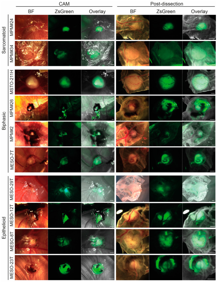
Figure 4.
MPM cell lines all form vascularised tumour nodules on the CAM. Representative images of tumour nodules formed by each of the 10 MPM cell lines for the experiment shown in Figure 3. Nodules are shown in situ on the CAM in ovo (left) and post-dissection viewing the nodule from beneath (right). Scale bar 1 mm. BF, bright field.

