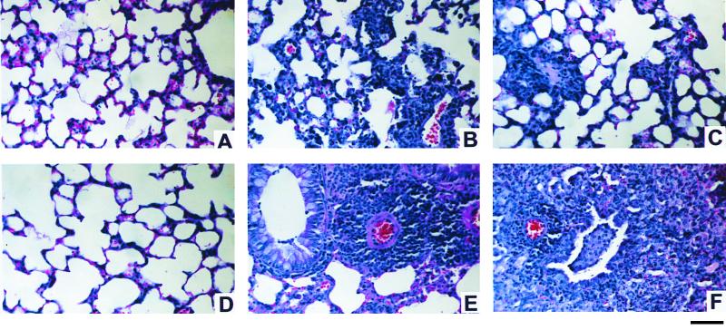
FIG. 1.
Histological comparison of lungs of mice at 7 days after C. pneumoniae infection or mock infection. All infected mice (B, C, E, and F) received 50 μl (2 × 10⁴ IFU) of C. pneumoniae intranasally. Anti-IL-12-treated mice (A and C) were injected with an IL-12-neutralizing MAb 1 day before infection and on day 5 p.i. Seven days after C. pneumoniae inoculation, lungs were harvested and prepared for histological study, as described in Materials and Methods. (A) Section from an anti-IL-12-treated uninfected BALB/c mouse, representing typical histology of uninfected BALB/c mice (treated with anti-IL-12 or control antibody). (B and C) Typical pneumonitis observed during infection in control antibody- and anti-IL-12-treated BALB/c mice, respectively. (D) Section from a G129 mouse, representing typical histology of uninfected wild-type 129 and G129 mice. (E) Typical perivascular cuffing in the lung of an infected 129 mouse. (F) Small vascular cuffing and bronchial infiltration in the lung of an infected G129 mouse. Bar, 100 μm.

