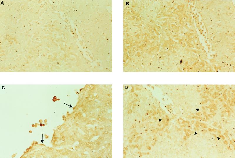
FIG. 6.
MIF liver immunohistochemistry of P. chabaudi-infected mice with acute disease (43% parasitemia). Panels A and B are serial sections: panel A is stained with preimmune serum, while panel B is stained with MIF antiserum (×20). Panel C shows immunoreactive inflammatory cells and endothelium (arrows) within the lumen of blood vessels (×40), while panel D illustrates MIF immunoreactivity in hepatocytes surrounding blood vessels (arrowheads) (×20).

