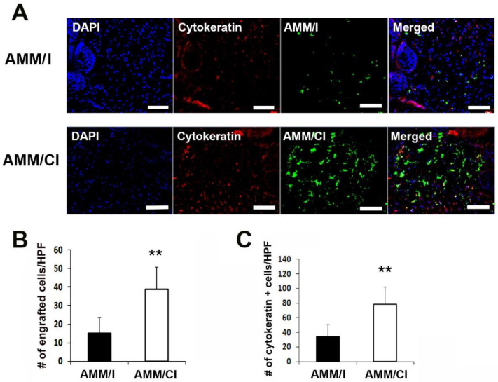
Figure 6.
AMM/CI showed high engraftment and epithelization potentials in wound tissue. (A) Representative images of cytokeratin immunostained wound tissue sections 10 days after cell injection. Bars = 200 μm. (B) Quantification of engrafted cells in the wound area after cell injection. ** p < 0.01, n = 7 per group. Engrafted AMM/CI were measured in the skin wound at 10 days after cell transplantation. (C) Quantification of cytokeratin-expressing cells in skin wound tissues. Cytokeratin-positive cells were measured in the skin wound tissues at 10 days after cell transplantation. ** p < 0.01, n = 6 per group.

