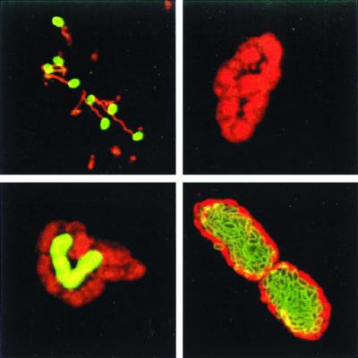
FIG. 1.
MAb 11A1 detects a differentially expressed spore wall antigen. HFF monolayers were infected with E. cuniculi spores and fixed 2 h (top left), 24 h (top right and bottom left), and 48 h (bottom right) postinfection. Double immunostaining was performed with MAb 11A1 detected with an FITC conjugate (green fluorescence), followed by staining with a polyclonal rabbit anti-microsporidia antiserum detected with a rhodamine conjugate (red fluorescence). MAb 11A1 stains a spore wall antigen located on the surfaces of extracellular spores. Expression of the spore wall antigen is first induced after 24 h in a small fraction of organisms located in the center of the vacuole. Most parasitophorous vacuoles contained MAb 11A1-positive E. cuniculi after 48 h. Note that a monolayer of negative organisms is located at the periphery of the vacuole.

