FIG. 2.
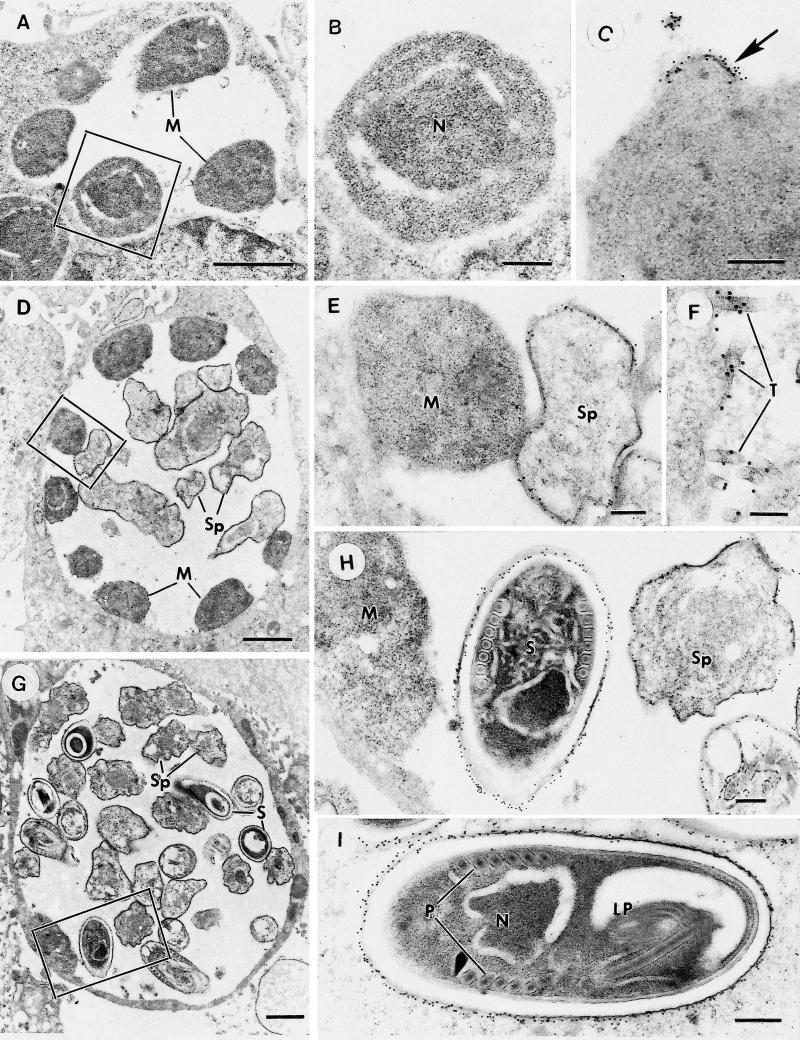
Immunoelectron microscopy of sections through cells infected with E. cuniculi at various stages of development which have been immunostained with MAb 11A1 and labeled with 10-nm gold particles. (A) Low-power micrograph of an early stage of intracellular development showing a parasitophorous vacuole containing peripherally located meronts (M). Bar, 1 μm. (B) Detail of the enclosed area in panel A. Note that the surface of the meront is unlabeled. Bar, 200 nm. (C) Part of a meront-like organism in which a focal area of the surface is strongly labeled (arrow). Note that the label is associated with an area of membrane thickening. Bar, 200 nm. (D) Low-power micrograph of a stage in development slightly later than that shown in panel A. The parasitophorous vacuole contains a few centrally located early sporonts (Sp) plus peripherally located meronts (M). Bar, 1 μm. (E) Enlargement of the enclosed area in panel D, contrasting the strong labeling of the surface of the early sporont (Sp) to the absence of staining of the adjacent meront (M). Bar, 200 nm. (F) Detail of the parasitophorous vacuole showing a number of labeled tubules (T) adjacent to the surface of a sporont. Bar, 100 nm. (G) Low-power micrograph of a cell containing E. cuniculi organisms at a late stage in development, in which the vacuole contains various stages in sporont development (Sp), including mature spores (S) with few remaining meronts. Bar, 1 μm. (H) Enlargement of the enclosed area in panel G, showing the absence of label on the meront (M) surface, while there is strong labeling of the surfaces of the sporont (Sp) and the mature spore (S). Bar, 200 nm. (I) Longitudinal section through a mature spore showing the strong labeling of the outer surface of the spore wall. N, nucleus; LP, lamellar polaroplast; P, polar tube. Bar, 200 nm.

