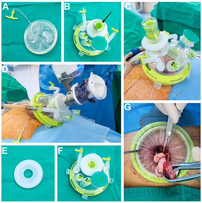
Figure 1.
(A) The wound retractor part of the Uni-port. (B) The cap part of the Uni-port with a laparoscopic port cap (black arrow) and four additional ports. (C) The assembled Uni-port for laparoscopic surgery. The CO2 line is connected, and abdominal cavity is inflated. (D) da Vinci® SP robot docking is completed. For robotic surgery, the port cap of the Uni-port is replaced, and the SP trocar is inserted. The CO2 line is connected, and the abdominal cavity is inflated. (E) The special port cap of the Uni-port for da Vinci® SP robot trocar. (F) The cap part of the Uni-port for the da Vinci® SP robot surgery system. The port cap indicated by the black arrow in Figure 1B has been replaced with the port cap for the da Vinci® SP robot trocar. (G) After the resected myoma is placed in an endopouch, it is pulled by hand, by holding it with a tenaculum forceps or towel clip. The inlet part of the endopouch is covered and fixed to the retractor part of the Uni-port. Subsequently, manual morcellation using scalpel is performed.

