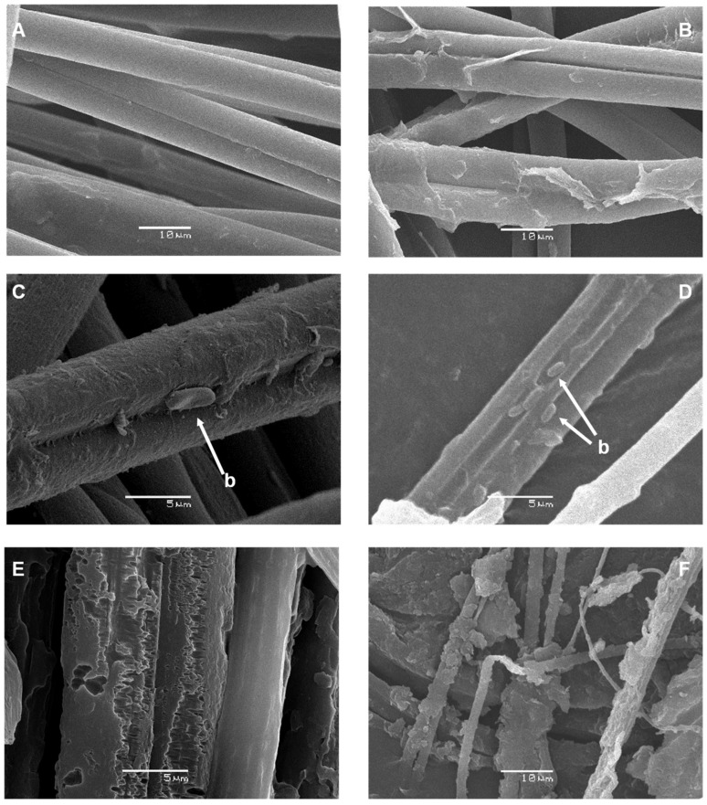
Figure 3.
Scanning electron microscopy (SEM) visualization of raw pine processionary silk filaments and their successive degradation over time mediated by a liquid culture of B. licheniformis. Silk native structure at 0 h of incubation (×2000) (A); partial removal of the sericin surface layer observed at 24 h of incubation (×2000) (B); almost complete elimination of the sericin layer detected at 36 h of incubation and presence of some bacterial cells (b, indicated by arrows) attached to the underlying fibroin fibers (×5000) (C) (×3000) (D); extensive degradation with loss of fibroin fiber structure observed after 48 h of incubation (×5000, (E)) (×2000, (F)).

