
FIGURE 1.
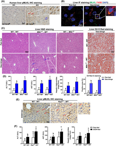
Differential roles of myeloid and nonmyeloid mixed lineage kinase domain‐like pseudokinase (Mlkl) and receptor interacting protein kinase 3 (Rip3) on Gao‐binge ethanol‐induced liver steatosis and injury. (A) Paraffin‐embedded human liver samples from healthy controls (HC; n = 10) and alcohol‐associated hepatitis (AH; n = 10) were stained for phospho‐MLKL (pMLKL; 40×) using standard immunohistochemistry (IHC) technique. Nuclei were counterstained with hematoxylin. pMLKL‐positive areas (%) were quantified using Image Pro‐Plus. pMLKL was localized in hepatocytes (blue arrows) and mononuclear immune cells (red arrows). (B) Double‐immunofluorescence (IF) staining for MLKL (green) and F4/80 (red) in murine liver from pair‐fed and Gao‐binge ethanol wild‐type (WT) mice. The magnified image illustrates colocalization (yellow). (C–F) Bone marrow chimeras were created as described in Materials and Methods and Figure S1A. Chimeras were allowed free access to Gao‐binge ethanol or pair‐fed control diets. (C) Hematoxylin and eosin (H&E) (10×) and Oil Red O (ORO; 20×) staining of livers from chimeric mice following the Gao‐binge protocol. n = 5–12 per group. (D) ALT and AST concentration in plasma, hepatic triglyceride content in whole liver homogenate, and quantification of ORO staining in Mlkl chimeras. (E) IHC staining for pMLKL (20×) in liver sections from WT→WT and Rip3 −/− →WT mice after ethanol (EtOH) feeding. Red arrows indicate positive staining in mononuclear immune cells. (F) ALT and AST concentration in plasma and hepatic triglyceride content in whole liver homogenate were detected in Rip3 chimeras, n = 3–7 per group. Values represent means ± SEM. Values with different superscripts are significantly different from each other. p < 0.05, assessed by t test (group = 2) or ANOVA (group ≥ 3).