
FIGURE 6.
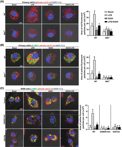
Effect of genetic and pharmacological inhibition of mixed lineage kinase domain‐like pseudokinase (MLKL) on phagocytosis by macrophages. (A,B) Primary mouse Kupffer cells (mKCs) isolated from C57BL/6J and Mlkl −/− mice were challenged with 10 ng/ml lipopolysaccharide (LPS), 50 mM ethanol (EtOH), or LPS + EtOH for 24 h following incubation with 50 μg/ml pHrodo red fluorescence conjugated Escherichia coli bioparticles for 3 h. Representative confocal images and quantification of (A) engulfed bioparticles and (B) particles colocalized with lysosomal associated membrane protein 1 (LAMP1); n = 4. (C) RAW cells were pretreated with GW806742X or GSK872 for 2 h and then exposed to LPS, EtOH, or LPS + EtOH for 24 h, as described above, followed by incubation with bioparticles for 3 h. Representative confocal images and quantification of colocalization with LAMP1 are shown. All images were obtained using a 63× objective (Zoom 4). Values represent means ± SEM. Values with different superscripts are significantly different from each other within the same color bars; n = 3, p < 0.05. WT, wild type. DIC, differential interference contrast.