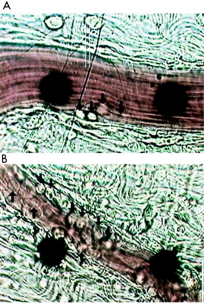
FIG. 4.
Photomicrographs of inflammation within the microvasculature following 3 h of treatment of rats with 4 × 10⁹ CFU of S. aureus M per kg. (A) S. aureus plus saline vehicle. (B) S. aureus plus 20 mg of imipenem per kg. Arrows point to leukocytes that have adhered to the endothelium. The black dots are part of an optical Doppler velocimeter used to measure centerline red blood cell velocity within the venule. Conditions of bacterial growth are as described in Materials and Methods.

