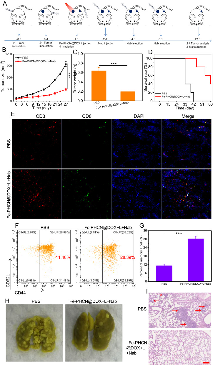
Fig. 9.
Abscopal effect and lung metastasis prevention in vivo. A Treatment schedule for Fe-PHCN@DOX and anti-PD-L1 nanobody combination therapy. Tumor growth curves (B) and tumor weights (C) from different groups. D Survival rates of mice after different treatments. E CLSM images of CD3+CD8+ T cells after staining with DAPI (blue), anti-CD3 antibody (red) and anti-CD8 antibody (green), respectively. The scale bar is 100 µm. The quantification (F) and percentage (G) of the effector memory T cells (CD3+CD8+CD44+CD62L−) by flow cytometric analyses after different treatments. H Representative images of the lung metastatic nodules. I H&E staining of lungs after different treatments. The scale bar is 200 μm. The p values were analyzed using the Log-rank (Mantel-Cox) test. Data are presented as the mean ± standard error of the mean. *p < 0.05, **p < 0.01, ***p < 0.001

