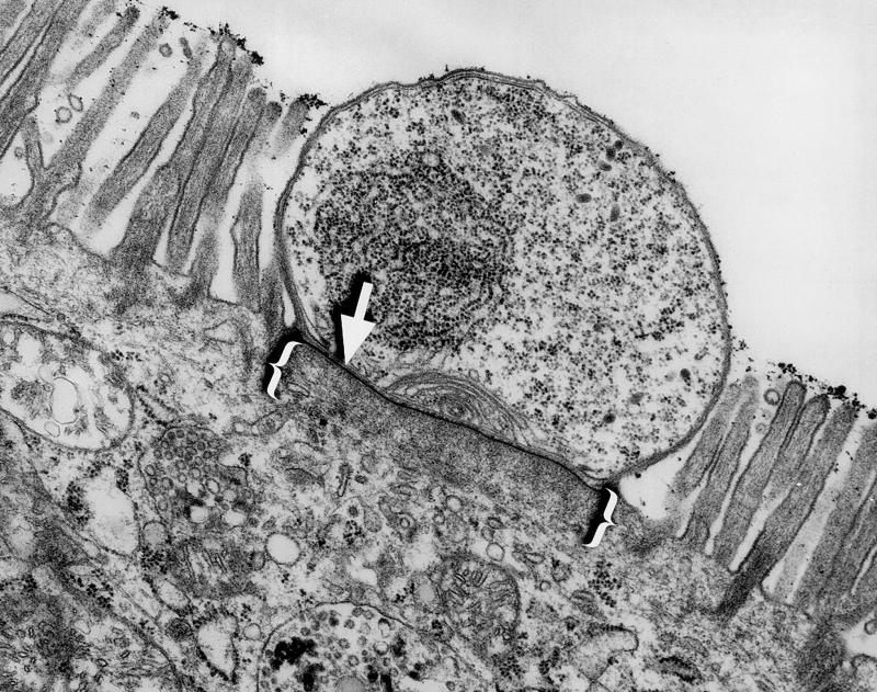
FIG. 1.
Transmission electron micrograph of a murine small intestinal epithelial cell infected by Cryptosporidium. Note the electron-dense band (arrow) separating the parasite from the host cell cytoplasm and the filamentous network immediately beneath this structure (brackets). Magnification, ×15,000.

