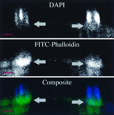
FIG. 3.
Confocal laser scanning microscopic longitudinal section through a DAPI- and FITC-phalloidin-stained Cryptosporidium-infected cell line demonstrating actin accumulation (arrows and green in composite) at the host-parasite interface, just below the parasite nuclei (blue in composite). The base of the host cell is toward the bottom of each photograph. Scale bars = 5 μm.

