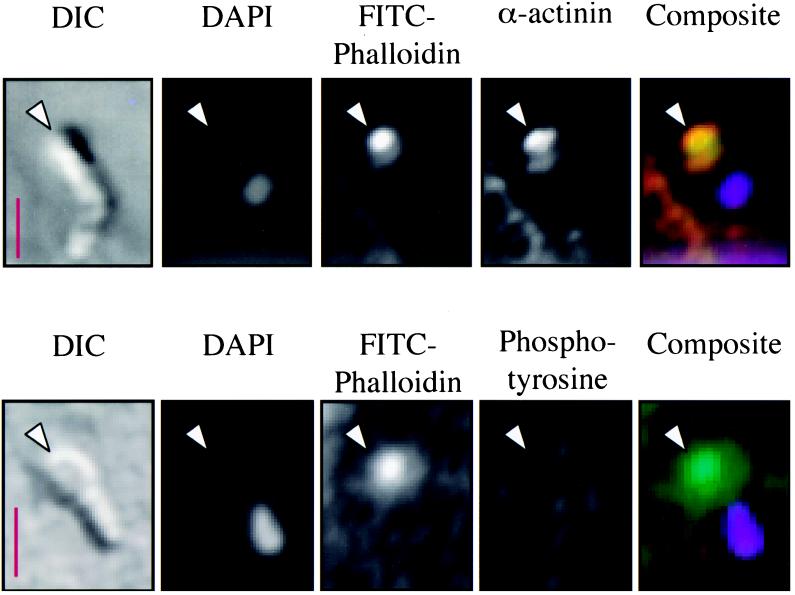
FIG. 4.
Fluorescence and DIC microscopy of two Cryptosporidium merozoites invading HCT-8 cells. The DIC images show elongated merozoites on the surface of the host cells, with the partially intracellular apical ends of the parasites indicated by arrowheads. The DAPI images of the same fields reveal the single merozoite nucleus toward the posterior end of the parasite. FITC-phalloidin stained f-actin is present at the site of invasion (arrowhead). Indirect immunofluorescence indicates the colocalization of α-actinin with f-actin early in invasion (yellow in composite, upper panel). The anterior portion of extracellular merozoites does not normally intensely express actin or α-actinin (data not shown). Also, there is no evidence of phosphotyrosine at this point of early invasion (lower panel). The basolateral focal adhesions, which are known to contain phosphotyrosine, were clearly positive in these cells (data not shown). Composite image: green, FITC-phalloidin; red, secondary antibody to α-actinin or phosphotyrosine; lavender, DAPI; yellow, colocalization of red and green. Scale bars = 2.5 μm.

