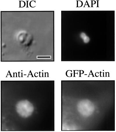
FIG. 5.
Fluorescence and DIC microscopy of a Cryptosporidium-infected MDCK cells constitutively expressing GFP-actin. The DIC image shows a Cryptosporidium meront in the MDCK cell line monolayer. The DAPI image of the same field reveals two nuclei of the parasite. Indirect immunofluorescence of the same field using antiactin antibodies highlights the actin plaque beneath the parasite. Fluorescence microscopy of the same field in the GFP fluorescence range reveals an image identical to the antiactin immunofluorescence, confirming the host cell origin of the actin plaque. Indirect immunofluorescence with anti-GFP antibodies produces identical staining of the actin plaque (data not shown). Scale bar = 5 μm.

